skip to main |
skip to sidebar
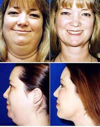
The use of barbed sutures appears to be a viable strategy for lifting and repositioning of facial tissue. The recently US FDA approved Contour Threads™ provides advantages to the surgeon and patient that other thread systems do not. However, the early success seen with Contour Thread™ lifts must stand the test of time.


Cosmetic Surgery, Breast Implant and Augmentation, Hair Transplant, Brow Lift,Rhinoplasty, Botox, Tummy Tuck
Aging of the face and neck results in ptosis of soft tissues and the appearance of more prominent facial lines.1 For correction of these changes, surgeons are increasingly reporting procedures with fewer incisions and shorter postoperative recovery periods. Many of these procedures utilize nonabsorbable sutures in the dermis and subcutis to lift lax skin.2-10 Limitations of these implants have included the protrusion of sutures through the skin,2-3 asymmetry of cosmetic effect requiring correction with additional sutures,3-5 and limited durability of effects.4 A new modified polypropylene suture marketed as Contour Threads™ was approved in 2005 by the US FDA for lifting ptotic skin of the face and neck. This implant is supplied as a 25cm polypropylene suture with a barbed middle portion. It is attached to a 7-inch straight needle distally and a 26mm 1/2 circle taper needle proximally.
Placement of Contour Threads™ to reduce ptosis of the brow, neck, middle and lower face may be performed in an outpatient setting under local anesthesia. The surgeon first establishes the degree and direction of the desired tightening. This determines the course and number of sutures that will be placed. Infiltration of local anesthesia is limited to these lines and the insertion points of the straight needle.
Because the Contour Thread™ barbs may be released with intense pressure, patients must initially avoid strenuous exercise or movements that could dislodge the tightened skin from the hundreds of barbs along the sutures. Non-peer reviewed data from the manufacturer demonstrate that in laboratory rats these sutures develop a fibrous capsule that becomes well integrated into the dermis and subcutaneous tissue over several months.11 Theoretically, a similar process in human skin could lead to a secure and long-lasting cosmetic effect. The actual long-term durability of the tightening effects of these sutures is unknown. Early adopters of this procedure have demonstrated maintenance of cosmetic effects at 6 months.12
The use of barbed sutures appears to be a viable strategy for lifting and repositioning of facial tissue. The recently US FDA approved Contour Threads™ provides advantages to the surgeon and patient that other thread systems do not. However, the early success seen with Contour Thread™ lifts must stand the test of time.
Subscribe to:
Post Comments (Atom)
Ear :Surgery
Hair And Head Surgery
Neck :Surgery
JOBS FOR INDIANS
Thigh Lift Surgery
Plastic Surgery Gone Wrong
Microdermabrasion
Blepharoplasty Surgery
Bad Plastic Surgery
Brow Lift Surgery
About Rhinoplasty
Mesotherapy
Facial Cosmetic Surgery
Facial Implants Surgery
Mini Face Lift Surgery
Fake Eyelashes Surgery
Botox
Facelift Surgery | Rhytidectomy information
Microdermabrasion Surgery
Dermabrasion Surgery
Injectable Fillers Surgery
Eyebrow Transplant
Eyebrow Restoration
Brow Lift
Eyelid Surgery or Blepharoplasty
Forehead Lifts OR Browplasty Surgery
Jaw Implant Surgery
Jaw Reduction Surgery
Jaw Augmentation Surgery
Scar Revision Surgery
Scar Removal Surgery
Cheek Implants Surgery
cheek augmentation surgery
Mentoplasty Surgery
Genioplasty Surgery
Chin Augmentation Surgery
Chin Implants Surgery
Nose Jobs / Rhinoplasty / Nose Surgery
Fat injections Surgery
Buccal fat extraction Surgery
Lip Reduction ( Cheiloplasty ) Surgery
Lip Augmentation / Lip Enhancement Surgery
Laser Skin Resurfacing Surgery
Mole Removal Surgery
Facial Liposuction Surgery
Buccal Fat Removal Surgery
Cheek Reduction Surgery
Mammoplasty or mammaplasty Surgery
Restylane Surgery
Intense Pulsed Light ( Photo Rejuvenation ) Surgery
Facial Rejuvenation surgery
Chemical Peels Surgery
Laser Freckles Removal Surgery
About Laser Warts Removal
QUESTIONSS OF LASER EYE SURGERY
Laser Hair Removal Surgery
Laser Tattoo Removal Surgery
Laser Scar Removal Surgery
Laser Skin Resurfacing Surgery
Nose Jobs / Rhinoplasty / Nose Surgery
Iris implants Surgery
Symmastia Surgery
Subcision Surgery
Polyalkylimide Fillers Surgery
Polylactic Acid Surgery
Calcium Hydroxylapatite Surgery
Dermal Fillers Surgery
Botulinum Toxin Surgery
Contour threads Surgery
Cosmetic camouflage Surgery
Cryolipolysis Surgery
Threadlift Surgery
Birthmark Removal Surgery
Facial Fat Grafting Surgery
Skin Tightening Surgery
Photo Rejuvenation Surgery
Collagen Injections Surgery
Hyaluronic Acid Surgery
Thermage surgery
Breast Surgery Procedures :
Breast Removal Surgery
Breast Implants / Boob Job Surgery
Brest Lift/Mastopexy Surgery
Reduction Mammoplasty/Breast Reduction Surgery
Correction Of Inverted Nipples Surgery
Breast Implant Revision Surgery
Breast Reconstruction Surgery
Nipple Reconstruction Surgery
Benelli Lift Surgery
Macrolane Surgery
Breast Asymmetry Correction Surgery
Male Chest Reconstruction Surgery
Male Breast Reduction or Gynecomastia Surgery
Breast Augmentation Surgery
Celebrity Plastic Surgery
Kelendria "Kelly" Rowland,Celebrity Nose Job
Kelendria "Kelly" Rowland Breast Implants Surgery
Farrah Leni Fawcett Plastic Surgery
Cindy Lou Hensley Plastic Surgery
American comedian Carrot Top Plastic Surgery
American singer Jessica Simpson Nose Job
Sex Symbol Jessica Alba Nose Job
Jessica Biel Celebrity Nose Nose Job
Leighton Meester Celebrity Nose Jobs
Lipstick Jungle star Kim Raver Nose Job
American singer and television personality Heidi Nose Job
American actress and singer-songwriter Demi Lovato Nose Job
American acter Camilla Belle Nose Job
Celebrity Salma Hayek Before After
Salma Hayek's Celebrity Nose Job
Paula Jones Celebrity Nose Job
American recording artist.Lady Gaga Before And After
American recording artist.Lady Gaga Plastic Surgery
American acter Angelina Jolie Breast Implants?
An American actres Angelina Jolie Chin Implant
The American acter Angelina Jolie Plastic Surgery Before And After
Angelina Jolie Plastic Surgery
Lil Kim Plastic Surgery
Jessica Plastic Surgery
Meg Ryan Plastic Surgery
Meg Ryan Plastic Surgery Disaster
Jennifer Grey Rhinoplasty
Lil Kim’s breast implants are leaking
Victoria Beckham breast implants Surgery
Jennifer Grey Nose Job - The Shocking Truth
Jennifer plastic surgery
Tyra Banks Plastic Surgery Before and after pictures
American acter Blake Lively Plastic Surgery
Tyra Banks's breasts real?
Tyra Banks Cosmetic Surgery
Blake Lively nose job plastic surgery.
Amerie Plastic Surgery
Candice Patricia Bergen plastic surgery
Ashlee Nicole Simpson surgery
Nadya Suleman Surgery
Jennifer Grey Aniston Nose
Jennifer Grey Nose plastic surgery
Stacy Ann Ferguson plsatic surgery
Fergie Plastic Surgery
Plastic Surgery Of Madonna
Rhinoplasty Plastic Surgery
Otoplasty
Most Popular celebritys
Actress Salma Hayek Nose Job
Salma Hayek breast plastic surgery
Salma Hayek Before And After Surgery
Adrianne Curry breast surgery
Megan Fox Surgery
Angelina Jolie Surgery
Linsday Lohan breast Surgery
Beyonce Plastic Surgery
Heidi Montag Surgery
Singer Mariah Carey Plastic Surgery
Actress Courtney Cox Plastic Surgery
Actress Jada Pinkett Smith Plastic Surgery
Actress Jennifer Aniston plastic surgery
Actress Jennifer Grey Nose Job
Victoria Beckham breast Surgery
Acter Britney Spears nose job
Fergie rhinoplasty Plastic Surgery
Actress Pamela Anderson plastic surgery
Actress Sharon Stone nose job
Actress Tara Reid Breast Surgery
Actress Winona Ryder nose job
American actress Blake Lively Plastic Surgery
American Rapper and Singer Lil Kim Plastic Surgery
American actress Angelina Jolie Nose Job
Angelina Jolie Before After Surgery
Followers
Copyright 2009. Human Surgeryes - WPBoxedTech Theme Design by Technology Tricks for Health Coupons.
Bloggerized by Free Blogger Template - Sponsored by Graphic ZONe and Technology Info
Bloggerized by Free Blogger Template - Sponsored by Graphic ZONe and Technology Info


0 comments:
Post a Comment