Rhinoplasty procedures
Rhinoplasty can be performed under a general anesthetic, sedation, or with local anesthetic. Initially, local anesthesia, which is a mixture of lidocaine and epinephrine, is injected to numb the area and temporarily reduce vascularity. There are two possible approaches to the nose: closed approach and open approach. In closed rhinoplasty, incisions are made inside the nostrils. In open rhinoplasty, also known as a Coronal Forehead Lift[5], an additional inconspicuous incision is made across the columella (the bit of skin that separates the nostrils). The surgeon first separates the skin and soft tissues of the nose from the underlying structures. The cartilage and bone are reshaped, and the incisions are sutured closed. Some surgeons use a stent or packing inside the nose, followed by tape or stent on the outside.
In some cases, the surgeon may shape a small piece of the patient's own cartilage or bone, as a graft, to strengthen or change the shape of the nose. Usually the cartilage is harvested from the septum. If there isn't enough septum cartilage, which can occur in revision rhinoplasty, cartilage can be harvested from the concha of the ear or the ribs. In the rare case where bone is required, it is harvested from the cranium, the hip, or the ribs. Sometimes a synthetic implant may be used to augment the bridge of the nose.
 The incisions for a rhinoplasty are hidden inside the nose, with the exception of a small incision across the base of the nose, depicted by the dotted line.

The incisions allow the surgeon to see the size and shape of the cartilages and bones on the inside of the nose, so that they can be altered.
Here, the scissors are pointing out the lower lateral cartilage (in blue), which is one of the cartilages that gives the tip of the nose its shape. The red line shows the location of the planned incision across the bottom of the nose.

Once the skin has been lifted from the bone and cartilage framework of the nose, often the first task is to remove a hump, if one is present. Part of the hump is made of bone, and part of the hump is cartilage.
In the photograph, the black line shows the desired profile. The nose is made of bone above the scalloped grey line and cartilage below that line. The part of the hump made of bone is shaded red, and the part of the hump made of cartilage is shaded blue.
 The soft cartilage of the hump is removed with a scalpel, and the bony hump is often removed with a chisel, shown at the top of this photograph. "Osteotome" is the medical term for a chisel. This photograph also shows the copper hammer that is used with the osteotome.
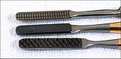 After the main part of the hump is removed with an osteotome, files are used to smooth out the remaining bone. The files are also called rasps, and they come in different shapes, orientations, and grades.
Some surgeons use rasps to remove the entire hump, foregoing use of the osteotome.
 One technique to narrow the nasal tip
A common complaint is that the tip of the nose is too wide. Many surgical techniques are available to narrow the tip of the nose, depending on what is causing the excess width.
In this photo, a suture is being placed to narrow the tip of the nose. The red line outlines the edge of the tip cartilage, which is narrowed when the suture tightens the fold of the cartilage at its apex. The suture is in light blue, ending in the needle, which appears white in the photograph. The cartilage is being held in place with tweezers, which are shaded green.
If the position of the nasal bones gives excess width to the upper part of the nose, the bones are moved inward, to a more narrow position. This skull shows in blue the position of the bones in the nose. For orientation, the eye sockets are outlined in red.

To narrow a nasal bone, two cuts are made in the bone with a tiny chisel: one cut starting at the yellow dot and extending up along the green arrow, and another cut starting at the blue dot and extending out along the black arrow. The piece of bone thus loosened from the skull is pushed inward, narrowing the nose.
These chisel cuts are made from underneath the skin, so there is no scar in the area after healing.

At the end of the procedure, after the incisions are closed, the nose is dressed, to hold it securely in place as it heals.
This photo shows the nose just before the dressing and splint are placed. The purple marks on the nose guided the surgeon in making accurate cuts in the bone during surgery.
 Preparing for the metal splint: the nose is first covered with paper tape in a manner to help maintain the nose's new shape.
 After taping, the metal splint is designed and cut and shaped, and it is placed on the nose.
 The metal splint is then covered with the tape, to hold it in place. The operation is now completed. The dressing will be removed in one week.
Revision rhinoplasty, also known as secondary rhinoplasty, is a nose operation performed to correct or revise an unsatisfactory outcome from a previous rhinoplasty. An unsatisfactory outcome occurs from 5% to 20% of rhinoplasties. There are two main reasons for performing secondary rhinoplasty. Patients often seek secondary rhinoplasty to correct a cosmetic deformity of the nose. A patient may be unsatisfied with all or part of a previous "nose reshaping.”. A nasal fracture may not have been reduced enough, or too much. A prominent or bulbous nasal tip may have not been addressed appropriately, or over-aggressively. The nose may looked pinched, it may look like a parrot’s beak, or like a boxer’s nose. There are many ways in which previous nose surgery may have left a nose aesthetically unappealing to a patient. The second reason is functional. The original nasal surgery may have been carried out to help with difficulties in breathing, and the outcome may have been unsatisfactory. Alternatively, the original surgery may have been performed for cosmetic reasons, but may have disrupted a normal physiologic mechanism involving the inspiration or expiration of air, making it difficult to breathe. Secondary rhinoplasty is a procedure often said to be extremely complicated. Because the nasal framework has often been destroyed or deformed from previous surgery, revision rhinoplasty experts frequently must reconstruct the support structures of the nose using cartilage grafts from either the ear (auricular cartilage graft) or from rib cartilage (costal cartilage graft). Most revision rhinoplasty specialists perform secondary rhinoplasty via the open approach. This allows the surgeon to directly visualize the deformity. Advances in rhinoplasty techniques, such as stabilization of rib cartilage grafts and utilization of the open approach, now allow satisfactory results in secondary rhinoplasty that were not possible in the past.
Rhinoplasty may be sought in the aftermath of traumatic deformity. Traumatic accidents are the most common cause of nasal deformity. Typically the nasal bones are broken and displaced. Occasionally, the nasal cartilages are disrupted or displaced, and in the worst cases the nasal dorsum is collapsed. Rhinoplasty allows shaving of the displaced bony humps, and re-alignment of the nasal bones after they are cut. When cartilage is disrupted, stitching of the cartilage for re-suspension, or use of cartilage grafts to camouflage depressions allows re-establishment of normal nasal contour. When the dorsum is collapsed, grafts of rib cartilage, ear cartilage, or cranial bone can be used to restore continuity to the dorsum. Although synthetic implants are also available for augmenting the nasal dorsum, cartilage or bone graft from the patient’s own body poses fewer risks of infection or rejection.[6]
 Rhinoplasty is sometimes sought for a collapsed nose due to septum perforation. Autoimmune problems such as Wegener’s Granulomatosis, Sarcoidosis, Churg-Strauss Syndrome, and Relapsing Polychondritis can lead to creation of a hole in the nasal septum, and loss of support in the dorsum leading to a saddle nose deformity. Intra nasal use of drugs such as cocaine, or extreme abuse of nasal decongestant sprays can similarly cause septum perforation and nasal dorsum collapse. Dorsum reconstruction is accomplished through the use of rib cartilage or bone grafts
Rhinoplasty to correct nasal obstruction following injudicious cosmetic surgery is common. Reconstructive rhinoplasty after injudicious cosmetic surgery allows the restoration of normal breathing. When nasal cartilages are over-aggressively trimmed during rhinoplasty, the nose can appear pinched and nasal potency compromised. Patients complain of nasal blockage that is worsened by attempts at deep inspiration. Internal cartilage grafts to support the nasal tip (batton grafts) or widen the middle vault of the nose (spreader grafts) can be quite effective in restoring normal breathing. These grafting techniques will increase the size of the nasal tip and widen the dorsum
Rhinoplasty for skin cancer excision also exists. Excision of skin cancers from the nose can lead to loss of internal support as well as external skin coverage. Skin cancer excision in the nose is commonly accomplished via the Mohs’ technique. Once the cancer is removed, reconstructive rhinoplasty aims to provide skin coverage utilizing techniques such as skin graft, local skin flaps, or pedicle flaps (see Nasal Reconstruction, Paramedian Forehead Flap). If cancer resection leads to loss of tissue in the area of the nasal tip, cartilage grafts are utilized to maintain support and prevent long-term distortion, by the force of scar contracture.
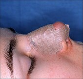
Rhinophyma is the late stage manifestation of a skin condition known as Rosacea, where the skin is infected with acne roseacea. The skin in the area of the nasal tip becomes red, thickened, and enlarged as exemplified by W C Fields. Although known acne treatments such as antibiotics and Acutane can halt the progression of this disease, thickening of the skin and obscuring of the nasal tip landmarks can only be remedied by surgical correction. Currently, laser excision of thickened abnormal skin represents the best option in rhinoplasty for Rhinophyma. The CO2 laser and the Erbium YAG laser are the most effective types of laser for this disorder
Vascular malformations and cleft lip anomalies are relatively common causes of congenital nasal deformities. In vascular malformations, the disease process can cause distortions of the skin and underlying structure of the nose. In cleft palate abnormalities, the size, position, and orientation of the nasal tip cartilages may be distorted. Rhinoplasty for reconstruction of vascular malformations can involve laser treatment of the skin and possible surgical excision. When the underlying cartilage structure is disturbed, cartilage grafts and stitching of the native nasal cartilages can help improve nasal appearance. In cleft lip patients, reconstructive rhinoplasty allows re-orientation of the nasal tip cartilages. Additional refinements with cartilage grafts to the tip are also frequently employed
Patients of African descent commonly seek narrowing of wide nostrils in a procedure known as alar base reduction. This procedure may include removing sections of the base of the nostrils or sections of the nose where it meets the face. Risk of keloid scar formation is very low, if the patient has not had keloids in the past. The tip of the nose can be restructured by removing tiny sections of cartilage to give the nose more definition, or adding cartilage grafts to provide additional structure to the nasal tip.
In some cases, the surgeon may shape a small piece of the patient's own cartilage or bone, as a graft, to strengthen or change the shape of the nose. Usually the cartilage is harvested from the septum. If there isn't enough septum cartilage, which can occur in revision rhinoplasty, cartilage can be harvested from the concha of the ear or the ribs. In the rare case where bone is required, it is harvested from the cranium, the hip, or the ribs. Sometimes a synthetic implant may be used to augment the bridge of the nose.
Skin incision for an open rhinoplasty. The incision may be “v-shaped” or a “stair-step” shaped incision. This aids the surgeon in attaining a precise closure and for camouflaging the resulting scar.
Exposing the cartilages inside the nose
The incisions allow the surgeon to see the size and shape of the cartilages and bones on the inside of the nose, so that they can be altered.
Here, the scissors are pointing out the lower lateral cartilage (in blue), which is one of the cartilages that gives the tip of the nose its shape. The red line shows the location of the planned incision across the bottom of the nose.
Planning excision of a nasal hump
Once the skin has been lifted from the bone and cartilage framework of the nose, often the first task is to remove a hump, if one is present. Part of the hump is made of bone, and part of the hump is cartilage.
In the photograph, the black line shows the desired profile. The nose is made of bone above the scalloped grey line and cartilage below that line. The part of the hump made of bone is shaded red, and the part of the hump made of cartilage is shaded blue.
Rhinoplasty osteotome and hammer
Rhinoplasty rasps
Some surgeons use rasps to remove the entire hump, foregoing use of the osteotome.
One technique to narrow the nasal tipA common complaint is that the tip of the nose is too wide. Many surgical techniques are available to narrow the tip of the nose, depending on what is causing the excess width.
In this photo, a suture is being placed to narrow the tip of the nose. The red line outlines the edge of the tip cartilage, which is narrowed when the suture tightens the fold of the cartilage at its apex. The suture is in light blue, ending in the needle, which appears white in the photograph. The cartilage is being held in place with tweezers, which are shaded green.
The nasal bones
Designing the cuts in the nasal bones
To narrow a nasal bone, two cuts are made in the bone with a tiny chisel: one cut starting at the yellow dot and extending up along the green arrow, and another cut starting at the blue dot and extending out along the black arrow. The piece of bone thus loosened from the skull is pushed inward, narrowing the nose.
These chisel cuts are made from underneath the skin, so there is no scar in the area after healing.
At the end of the rhinoplasty
At the end of the procedure, after the incisions are closed, the nose is dressed, to hold it securely in place as it heals.
This photo shows the nose just before the dressing and splint are placed. The purple marks on the nose guided the surgeon in making accurate cuts in the bone during surgery.
Taping the nose, in preparation for the metal splint
Metal nasal splint in place
Metal nasal splint has been taped on the nose
Primary and secondary
Primary rhinoplasty refers to first-time rhinoplasty whether it is performed for aesthetic, functional, or reconstructive purposes.Revision rhinoplasty, also known as secondary rhinoplasty, is a nose operation performed to correct or revise an unsatisfactory outcome from a previous rhinoplasty. An unsatisfactory outcome occurs from 5% to 20% of rhinoplasties. There are two main reasons for performing secondary rhinoplasty. Patients often seek secondary rhinoplasty to correct a cosmetic deformity of the nose. A patient may be unsatisfied with all or part of a previous "nose reshaping.”. A nasal fracture may not have been reduced enough, or too much. A prominent or bulbous nasal tip may have not been addressed appropriately, or over-aggressively. The nose may looked pinched, it may look like a parrot’s beak, or like a boxer’s nose. There are many ways in which previous nose surgery may have left a nose aesthetically unappealing to a patient. The second reason is functional. The original nasal surgery may have been carried out to help with difficulties in breathing, and the outcome may have been unsatisfactory. Alternatively, the original surgery may have been performed for cosmetic reasons, but may have disrupted a normal physiologic mechanism involving the inspiration or expiration of air, making it difficult to breathe. Secondary rhinoplasty is a procedure often said to be extremely complicated. Because the nasal framework has often been destroyed or deformed from previous surgery, revision rhinoplasty experts frequently must reconstruct the support structures of the nose using cartilage grafts from either the ear (auricular cartilage graft) or from rib cartilage (costal cartilage graft). Most revision rhinoplasty specialists perform secondary rhinoplasty via the open approach. This allows the surgeon to directly visualize the deformity. Advances in rhinoplasty techniques, such as stabilization of rib cartilage grafts and utilization of the open approach, now allow satisfactory results in secondary rhinoplasty that were not possible in the past.
Functional and reconstructive
Reconstructive rhinoplasty refers to restoring the normal shape and function of the nose following damage from a traumatic accident, autoimmune disorder, intra-nasal drug abuse, previous injudicious cosmetic surgery, cancer involvement, or congenital abnormality. Rhinoplasty can restore skin coverage, recreate normal contours, and re-establish nasal airflow. To improve nasal breathing function, a septoplasty may also be performed. If there is turbinate hypertrophy, an inferior turbinectomy can be performed.Rhinoplasty may be sought in the aftermath of traumatic deformity. Traumatic accidents are the most common cause of nasal deformity. Typically the nasal bones are broken and displaced. Occasionally, the nasal cartilages are disrupted or displaced, and in the worst cases the nasal dorsum is collapsed. Rhinoplasty allows shaving of the displaced bony humps, and re-alignment of the nasal bones after they are cut. When cartilage is disrupted, stitching of the cartilage for re-suspension, or use of cartilage grafts to camouflage depressions allows re-establishment of normal nasal contour. When the dorsum is collapsed, grafts of rib cartilage, ear cartilage, or cranial bone can be used to restore continuity to the dorsum. Although synthetic implants are also available for augmenting the nasal dorsum, cartilage or bone graft from the patient’s own body poses fewer risks of infection or rejection.[6]
The lower lateral cartilage (greater alar cartilage) exposed through the left nostril for modification during a rhinoplasty.
Rhinoplasty to correct nasal obstruction following injudicious cosmetic surgery is common. Reconstructive rhinoplasty after injudicious cosmetic surgery allows the restoration of normal breathing. When nasal cartilages are over-aggressively trimmed during rhinoplasty, the nose can appear pinched and nasal potency compromised. Patients complain of nasal blockage that is worsened by attempts at deep inspiration. Internal cartilage grafts to support the nasal tip (batton grafts) or widen the middle vault of the nose (spreader grafts) can be quite effective in restoring normal breathing. These grafting techniques will increase the size of the nasal tip and widen the dorsum
Rhinoplasty for skin cancer excision also exists. Excision of skin cancers from the nose can lead to loss of internal support as well as external skin coverage. Skin cancer excision in the nose is commonly accomplished via the Mohs’ technique. Once the cancer is removed, reconstructive rhinoplasty aims to provide skin coverage utilizing techniques such as skin graft, local skin flaps, or pedicle flaps (see Nasal Reconstruction, Paramedian Forehead Flap). If cancer resection leads to loss of tissue in the area of the nasal tip, cartilage grafts are utilized to maintain support and prevent long-term distortion, by the force of scar contracture.
Rhinophyma is the late stage manifestation of a skin condition known as Rosacea, where the skin is infected with acne roseacea. The skin in the area of the nasal tip becomes red, thickened, and enlarged as exemplified by W C Fields. Although known acne treatments such as antibiotics and Acutane can halt the progression of this disease, thickening of the skin and obscuring of the nasal tip landmarks can only be remedied by surgical correction. Currently, laser excision of thickened abnormal skin represents the best option in rhinoplasty for Rhinophyma. The CO2 laser and the Erbium YAG laser are the most effective types of laser for this disorder
Vascular malformations and cleft lip anomalies are relatively common causes of congenital nasal deformities. In vascular malformations, the disease process can cause distortions of the skin and underlying structure of the nose. In cleft palate abnormalities, the size, position, and orientation of the nasal tip cartilages may be distorted. Rhinoplasty for reconstruction of vascular malformations can involve laser treatment of the skin and possible surgical excision. When the underlying cartilage structure is disturbed, cartilage grafts and stitching of the native nasal cartilages can help improve nasal appearance. In cleft lip patients, reconstructive rhinoplasty allows re-orientation of the nasal tip cartilages. Additional refinements with cartilage grafts to the tip are also frequently employed
Ethnic
Although techniques and methods employed during rhinoplasty surgeries are the same regardless of ethnicity[citation needed], there are some trends that apply to patients of certain ethnic backgrounds, due to their similar anatomic features. East Asian patients often want their noses to appear narrower and their bridges higher. If very little elevation of the bridge is desired, the nasal bones can be cut and moved towards the midline. This technique will narrow the bridge and also cause a slight elevation in the dorsum. East Asian patients who seek greater augmentation of the bridge of their nose require implants. A variety of alloplastic implants including Gore-Tex, Med-Por, or silicone can be used. Tissues from the patient's own body (autologous) can be used for augmentation, in order to reduce the risk of complications such as infection or extrusion. Septum cartilage, rib cartilage (costal cartilage), ear cartilage (auricular cartilage), and fascia are being often used. In non surgical rhinoplasty, filler materials such as hyaluronic acid or calcium based microspheres can be injected under the skin, in the bridge of the nose. These injections however, are non permanent lasting between six months to a year.Patients of African descent commonly seek narrowing of wide nostrils in a procedure known as alar base reduction. This procedure may include removing sections of the base of the nostrils or sections of the nose where it meets the face. Risk of keloid scar formation is very low, if the patient has not had keloids in the past. The tip of the nose can be restructured by removing tiny sections of cartilage to give the nose more definition, or adding cartilage grafts to provide additional structure to the nasal tip.


0 comments:
Post a Comment